




p (1).png)




.png)




.png)

Case Report
Concurrent Nutcracker Syndrome and Median Arcuate Ligament Syndrome: A Case Report
- Dr. Eman Jamal Saeed Shawahna
Corresponding author: Eman Jamal Saeed Shawahna, Department: Clinical research, An-Najah national university, Nablus, Palestine, ORCID ID: 0009-0002-4746-3371.
Volume: 3
Issue: 2
Article Information
Article Type : Case Report
Citation : Eman Jamal Saeed Shawahna, Alaa Atef Mohmmad Hasan. Concurrent Nutcracker Syndrome and Median Arcuate Ligament Syndrome: A Case Report. Journal of Medical and Clinical Case Reports 3(2). https://doi.org/10.61615/JMCCR/2026/JULY027140722
Copyright: © 2026 Eman Jamal Saeed Shawahna. This is an open-access article distributed under the terms of the Creative Commons Attribution License, which permits unrestricted use, distribution, and reproduction in any medium, provided the original author and source are credited.
DOI: https://doi.org/10.61615/JMCCR/2026/JULY027140722
Publication History
Received Date
20 Jun ,2026
Accepted Date
13 Jul ,2026
Published Date
22 Jul ,2026
Abstract
Introduction
Vascular compression syndromes are a rare disorder, though late detection is common. However, significant clinical consequences occur. The concurrent presence of two of these syndromes is rare. Proper diagnosis and management are mandatory to avoid missing such cases.
Case Presentation
A thirty-year-old male patient with a free past medical and surgical history experienced sudden weight loss due to psychological distress. After that, he started to complain of abdominal pain for 8 months. The dominant symptom was postprandial abdominal pain, which exacerbated his weight loss. He underwent CT angiography, which revealed a coexistence of median arcuate ligament syndrome and nutcracker syndrome. He underwent surgical management with an excellent outcome.
Conclusion
Doppler ultrasound is not always sensitive for diagnosing vascular compression syndromes, so further evaluation with CT angiography should be pursued when symptoms cannot be explained by Doppler ultrasound and there remains a high suspicion of a vascular syndrome. Surgical intervention is the gold standard treatment for median arcuate ligament syndrome. When incidental findings on NCS occur, follow-up with urine analysis may be considered a simple, accessible test. Treatment to prevent thrombosis could also be considered even in asymptomatic patients. Instructions should be given to the patient about symptoms that could indicate or be associated with NCS, to facilitate follow-up and adjust the plan as needed when the syndrome becomes symptomatic. Additionally, guidance on GI obstruction and Wilkie syndrome should be provided.
►Concurrent Nutcracker Syndrome and Median Arcuate Ligament Syndrome: A Case Report
Eman Jamal Saeed Shawahna1*, Alaa Atef Mohmmad Hasan2
1Department: Clinical research, An-Najah national university, Nablus, Palestine, ORCID ID: 0009-0002-4746-3371.
2Department: Faculty of medicine, An-Najah national university, Nablus, Palestine.
Introduction
Abdominal vascular compression syndromes (AVCS) encompass a group of clinical conditions caused by extrinsic compression of blood vessels by adjacent anatomical structures, or compression of hollow viscera by neighboring vessels. Although individually uncommon, these syndromes, including nutcracker syndrome (NCS), median arcuate ligament syndrome (MALS), May-Thurner syndrome, and superior mesenteric artery syndrome, can produce significant morbidity when unrecognized. Their nonspecific symptomatology and rarity frequently lead to delayed diagnosis, and the coexistence of two or more vascular compression syndromes in a single patient is exceedingly rare [1-5].
Nutcracker syndrome refers to symptomatic compression of the left renal vein (LRV), most commonly at the level of the superior mesenteric artery and the abdominal aorta (anterior NCS). However, posterior entrapment behind the aorta has also been described. The distinction between the nutcracker phenomenon, which is an anatomical finding of LRV compression without clinical manifestations, and nutcracker syndrome, in which symptoms are present, is clinically important. When symptomatic, NCS may manifest with hematuria, left flank pain, orthostatic proteinuria, pelvic congestion symptoms in women (dyspareunia, dysmenorrhea, chronic pelvic pain), and varicocele in men. The etiology may be congenital or acquired, and significant complications include chronic kidney disease from sustained renal venous hypertension and LRV thrombosis. Diagnosis relies on a stepwise approach: Doppler ultrasonography, followed by computed tomography angiography (CTA) or magnetic resonance angiography (MRA), with retrograde venography and renocaval pressure gradient measurement serving as confirmatory tools. Management ranges from conservative observation to open, laparoscopic, or endovascular surgical interventions, depending on symptom severity [6-9].
Median arcuate ligament syndrome is a rare vascular compression syndrome with an estimated incidence of 2 per 100,000 individuals, characterized by extrinsic compression of the celiac artery by the median arcuate ligament and diaphragmatic crura. The pathophysiology remains debated; one widely accepted theory proposes that compression of the celiac artery leads to foregut ischemia during periods of increased blood demand, while an alternative hypothesis implicates neuropathic pain from chronic compression and overstimulation of the celiac ganglion. MALS is more prevalent in women and in individuals with a thin body habitus, and typically presents with postprandial epigastric pain, nausea, vomiting, and unintentional weight loss. It remains a diagnosis of exclusion, as celiac artery compression is a common incidental finding in asymptomatic individuals. Duplex ultrasonography serves as a reasonable initial imaging modality, while CT angiography provides detailed anatomical delineation of the compressed celiac artery and the position of the median arcuate ligament. The definitive treatment is surgical decompression of the celiac artery, with celiac ganglion neurolysis considered for neuropathic pain relief [10-12].
The coexistence of NCS and MALS in a single patient has rarely been reported in the literature. Linares et al. described the first documented case of concurrent Dunbar (MALS) and nutcracker syndromes, noting that while no etiologic relationship exists between the two entities, their co-occurrence poses unique diagnostic and therapeutic challenges [5]. Weight loss, which may be both a consequence of MALS-related food avoidance and a contributing factor to the unmasking of vascular compression due to loss of retroperitoneal fat, can create a vicious cycle that perpetuates both conditions [12].
Case Presentation
A thirty-year-old male patient, with no significant past medical or surgical history, married and having six healthy siblings, began experiencing abdominal pain 8 months ago following a sudden weight loss caused by emotional factors. The pain initially localized in the lower abdomen and later migrated to the epigastric region after meals, accompanied by bloating and postprandial stomach fullness, despite having a good appetite and feeling hungry. However, he couldn’t eat due to postprandial abdominal pain. He reported no diarrhea, fever, or vomiting. The patient developed an eating phobia, which worsened his weight loss due to postprandial pain, and he also experienced constipation and an anal fissure.
History
Free of past medical and surgical history. No known drug or food allergies, no use of illicit or licit drugs, and no alcohol consumption. He has a history of H. pylori infection diagnosed by breath test, which was treated with no symptom improvement.
Diagnostic Assessment
A comprehensive laboratory workup was conducted. Complete blood count, liver function tests, renal function tests, and thyroid function tests all returned normal results. Urinalysis was normal, with no signs of hematuria or proteinuria.
Initial abdominal ultrasonography showed no abnormalities, with no signs of gallstones, hepatobiliary issues, or other structural problems. Doppler ultrasonography was performed twice; neither exam detected hemodynamically significant celiac artery compression or left renal vein entrapment.
An upper gastrointestinal endoscopy and colonoscopy were performed to evaluate mucosal pathology. Both exams were unremarkable, showing no signs of ulceration, inflammation, or neoplasia.
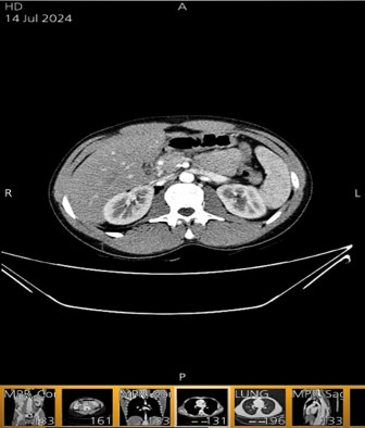
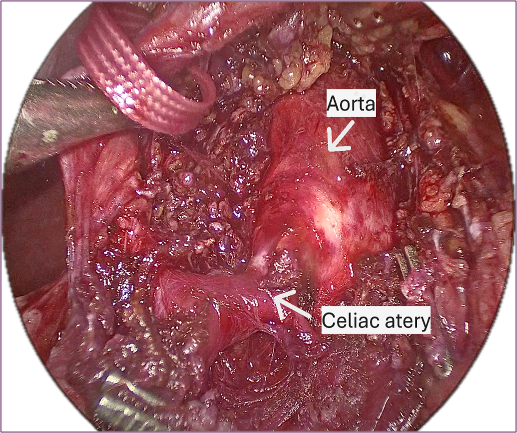
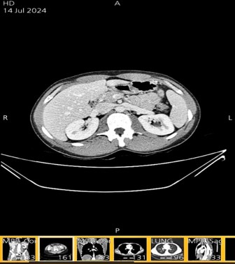
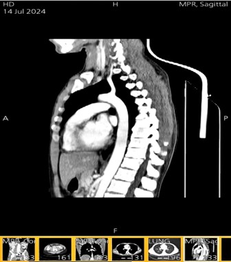
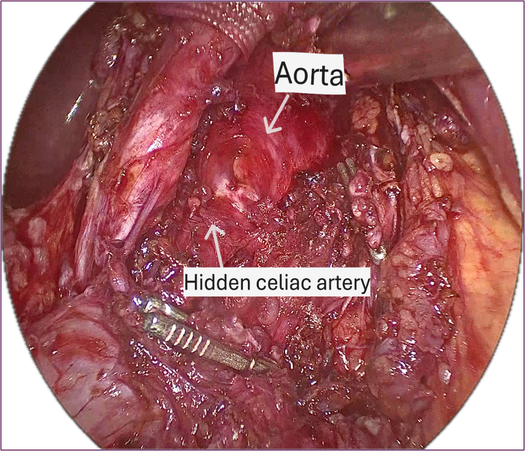
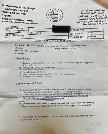
Given the ongoing symptoms despite a thorough negative workup and a high clinical suspicion of a vascular cause, computed tomography angiography (CTA) of the abdomen was performed. CTA showed two simultaneous findings: compression of the celiac artery by the median arcuate ligament, which indicates median arcuate ligament syndrome (MALS), and compression of the left renal vein between the superior mesenteric artery and the abdominal aorta, consistent with the nutcracker phenomenon. No signs of superior mesenteric artery syndrome (Wilkie syndrome) or other vascular compression were found.
Intervention
The patient was afraid of surgery and inquired about an alternative plan. He considered weight regain as a potential solution to his problem, but it was impractical due to postprandial pain and limited food intake. Ultimately, after discussing options with his internist, he decided to proceed with surgery as a definitive treatment to end his suffering and return to his normal daily life.
Follow-up and patient perspective on intervention
In the first few days after surgery, the patient experienced constitutional side effects of abdominal surgery, including nausea, bloating, and mild abdominal pain. Two months post-surgery, the patient expressed great gratitude, showing significant progress and satisfactory improvement in postprandial abdominal pain. He started eating as normally as he did before his sudden weight loss and the development of median arcuate ligament syndrome symptoms. He described his improvement as follows: (It’s like magic; I am so thankful for diagnosing me and advising me to undergo the surgery; you brought my life back, thank you very much for being my doctor). After full recovery, instructions to monitor for nutcracker syndrome symptoms and to undergo periodic urinalysis tests were advised.
Informed Consent
The patient agreed and was allowed to report and publish the case, and the purpose of reporting the case was clearly explained to the patient.
Discussion
This case illustrates several important clinical notes regarding the diagnosis and management of concurrent abdominal vascular compression syndromes.
Diagnostic Challenges and Delayed Recognition
Abdominal vascular compression syndromes are frequently underdiagnosed due to their rarity, nonspecific symptomatology, and clinicians' limited familiarity with these conditions. [1,2]. The clinical presentation of MALS, including epigastric pain, postprandial discomfort, nausea, and weight loss, overlaps substantially with far more common gastrointestinal disorders, leading to extensive and often unrevealing diagnostic workups before the correct diagnosis is considered. In the present case, the patient underwent upper and lower endoscopy, abdominal ultrasonography, and comprehensive laboratory investigations (all of which were unremarkable) before CT angiography ultimately revealed both left renal vein compression and celiac artery compression by the median arcuate ligament. This diagnostic trajectory is consistent with the literature, which reports that patients with MALS typically undergo prolonged evaluation by primary care physicians and gastroenterologists before referral to a vascular specialist [13]. Similarly, the diagnosis of NCS is commonly delayed because its symptoms, particularly flank pain and nonspecific abdominal discomfort, are attributed to more prevalent conditions [8].
The distinction between the nutcracker phenomenon and nutcracker syndrome is clinically significant because treatment depends on it. The nutcracker phenomenon describes an anatomical finding of left renal vein compression without symptoms, while nutcracker syndrome involves clinical signs such as hematuria, flank pain, orthostatic proteinuria, or varicocele [8-14].
In the present case, the left renal vein compression was identified incidentally on CTA performed primarily to evaluate the patient's gastrointestinal symptoms, and the patient did not exhibit classic renal manifestations of NCS such as hematuria or proteinuria. This finding, therefore, represents a nutcracker phenomenon at the time of presentation rather than a nutcracker syndrome. Nevertheless, the incidental discovery of left renal vein compression warrants monitoring, as undiagnosed NCS may progress to significant morbidity, including chronic kidney disease from sustained renal venous hypertension and left renal vein thrombosis [7]. Periodic urinalysis represents a simple, cost-effective surveillance tool for detecting the transition from an asymptomatic phenomenon to a symptomatic syndrome. In pediatric cohorts, conservative management with longitudinal follow-up has demonstrated spontaneous resolution of hematuria and proteinuria in approximately 40% of patients within three years, supporting an initial observation strategy [15,16].
Limitations of Doppler Ultrasonography
A notable finding in this case was the failure of Doppler ultrasonography (DUS), performed on two separate occasions, to identify either MALS or NCS. While DUS is recommended as a reasonable initial screening modality for both conditions due to its cost-effectiveness, lack of ionizing radiation, and ability to provide hemodynamic information, it has recognized limitations. For MALS, DUS can demonstrate elevated celiac artery velocities during expiration, but its sensitivity is operator-dependent and may miss compression in certain anatomic configurations [6,12]. For NCS, the sensitivity of color Doppler sonography has been reported at 78% with a specificity of 100% when collateral venous flow is included in the diagnostic criteria. However, technical factors, including Doppler angle, sample volume, patient body habitus, and operator experience, can significantly affect diagnostic accuracy [17,18].
In contrast, CT angiography provides superior anatomical detail with three-dimensional reconstruction capabilities, allowing simultaneous evaluation of multiple vascular territories and identification of concomitant abdominal pathology. The American College of Radiology Appropriateness Criteria recommend CTA as the initial imaging study for suspected mesenteric ischemia, noting sensitivity and specificity of 95–100% for grading mesenteric vessel stenosis, and specifically recognizing CTA as an accurate tool for detecting MALS [1,3,19]. Multidetector CT is particularly valuable in AVCS because it allows a comprehensive single-study evaluation of the anatomy and resultant morphologic changes across multiple vascular compression syndromes simultaneously [1]. This case underscores that when clinical suspicion for a vascular compression syndrome remains high despite negative DUS, further evaluation with CTA should be pursued.
The Role of Weight Loss: A Vicious Cycle
Weight loss plays a dual role in the pathophysiology of AVCS, functioning as both a consequence and a perpetuating factor. MALS is more prevalent in individuals with a thin body habitus, and loss of retroperitoneal and mesenteric fat may reduce the cushioning effect around the celiac axis, exacerbating compression. Simultaneously, the postprandial abdominal pain characteristic of MALS leads to food avoidance (sitophobia) and progressive weight loss, creating a self-reinforcing cycle. In the present case, the patient's initial weight loss that has been attributed to emotional factors may have unmasked or worsened the celiac artery compression, which in turn caused postprandial pain and eating phobia, leading to further weight loss. This vicious cycle is well described in chronic mesenteric ischemia, where malnutrition at the time of presentation has been associated with increased perioperative mortality and decreased long-term survival after revascularization [12,13].
Similarly, loss of retroperitoneal fat may contribute to narrowing of the aortomesenteric angle, potentially exacerbating compression of the left renal vein. The coexistence of SMA syndrome (Wilkie syndrome) with NCS has been reported in patients with significant weight loss, as both conditions share an anatomic relationship between the superior mesenteric artery and the aorta [20]. Although the present patient did not demonstrate duodenal compression, the shared anatomic predisposition highlights the importance of evaluating for multiple compression syndromes when one is identified, particularly in the setting of significant weight loss.
Coexistence of MALS and NCS
The concurrent presentation of MALS and NCS in a single patient is exceedingly rare. Linares et al. described the first documented case of this association, noting that no etiologic relationship exists between the two entities, as they involve compression of different vascular structures by distinct anatomic mechanisms. However, both conditions may be unmasked or exacerbated by the same predisposing factors, particularly thin body habitus and loss of retroperitoneal fat, which may explain their co-occurrence [5,20]. The present case adds to this limited body of literature and reinforces the importance of comprehensive vascular imaging when an AVCS is identified, as additional compression syndromes may coexist.
Recent literature has also highlighted an association between multiple AVCS and connective tissue disorders, particularly hypermobile Ehlers-Danlos syndrome (hEDS) and hypermobility spectrum disorder, in which the prevalence of MALS, NCS, May-Thurner syndrome, and SMA syndrome appears to be increased [4,12]. Although the present patient was not evaluated for connective tissue disorders, this association may warrant consideration in patients presenting with multiple concurrent AVCS.
Management Considerations
The management of concurrent MALS and NCS presents unique challenges. For MALS, the definitive treatment is surgical decompression of the celiac artery through division of the median arcuate ligament and diaphragmatic crura, with celiac ganglion neurolysis considered for neuropathic pain relief [10,12]. Surgical decompression has demonstrated symptom improvement in approximately 75–85% of patients in the short to medium term, though long-term data beyond five years remain limited [10,22]. Outcome predictors include postexertional pain (favorable) and unprovoked pain or vomiting (unfavorable). Patients treated nonoperatively appear to have worse outcomes, with only 4% achieving complete symptom resolution compared with 37% in the surgical group in one series [23].
Conservative management, including counseling, analgesia, and dietary modifications, has been described for MALS, but prospective comparative data are lacking [24]. Weight regain, while theoretically beneficial by increasing retroperitoneal fat and reducing celiac compression, is often impractical in patients with MALS because the postprandial pain that drives weight loss persists until the underlying compression is addressed [13]. This paradox was clearly demonstrated in the present case, where the patient was unable to regain weight due to ongoing ischemic abdominal pain and food avoidance.
For the incidentally discovered nutcracker phenomenon, conservative management with periodic surveillance is appropriate in the absence of symptoms. Urinalysis should be performed at regular intervals to monitor for the development of hematuria or proteinuria, which would signal progression to nutcracker syndrome. Patient education regarding symptoms that may indicate progression, including hematuria, worsening flank pain, or pelvic congestion symptoms, is essential to guide timely reassessment and potential escalation of management [7,16]. If NCS becomes symptomatic and refractory to conservative measures, treatment options include left renal vein transposition, renal autotransplantation, extravascular stenting, and endovascular stenting, with reported symptom resolution rates ranging from 52% with conservative management to 92% with LRV transposition [25].
Conclusion
Doppler ultrasound is not always sensitive for diagnosing vascular compression syndromes, so further evaluation with CT angiography should be pursued when symptoms cannot be explained by Doppler ultrasound and there remains a high suspicion of a vascular syndrome. Surgical intervention is the gold standard treatment for median arcuate ligament syndrome. When incidental findings on NCS occur, follow-up with urine analysis may be considered a simple, accessible test. Treatment to prevent thrombosis could also be considered even in asymptomatic patients. Instructions should be given to the patient about symptoms that could indicate or be associated with NCS, to facilitate follow-up and adjust the plan as needed when the syndrome becomes symptomatic. Additionally, guidance on GI obstruction and Wilkie syndrome should be provided.
- Lamba R, Tanner DT, Sekhon S, McGahan JP, Corwin MT, Lall CG. (2014). Multidetector CT of vascular compression syndromes in the abdomen and pelvis. Radiographics. 34(1):93-115.
- Nadim B, Alizada S, Gupta S, Steigner M, Menard M, Aghayev A. (2024). Under pressure: a head-to-toe review of vascular compression syndromes. Clinical Radiology. 79(10):722-35.
- Cardoso DL, de Macedo BA, Neto RM, Cardoso MTL, Marciano L, de Farias LdPG, (2025). Abdominal vascular compression syndromes: A pictorial review. European Journal of Radiology. 189:112169.
- Bruessel P, Govender M, Frahm-Jensen G. (2026). The Investigation and Management of the Abdominopelvic Vascular Compression Syndromes in Patients with Ehlers–Danlos Syndrome and Hypermobility Spectrum Disorder. Vascular Health and Risk Management. 592420.
- Linares P, Vivas S, Dominguez A, Jorquera F, Muñoz F, Espinel J, (2002). An uncommon association of abdominal vascular compression syndromes: Dumbar and Nutcracker. European journal of gastroenterology & hepatology. 14(10):1151-3.
- Orczyk K, Wysiadecki G, Majos A, Stefańczyk L, Topol M, Polguj M. (2017). What each clinical anatomist has to know about left renal vein entrapment syndrome (nutcracker syndrome): a review of the most important findings. BioMed Research International. (1):1746570.
- Ananthan K, Onida S, Davies A. (2017). Nutcracker syndrome: an update on current diagnostic criteria and management guidelines. European journal of vascular and endovascular surgery. 53(6):886-94.
- Kurklinsky AK, Rooke TW, editors. (2010). Nutcracker phenomenon and nutcracker syndrome. Mayo Clinic Proceedings. Elsevier.
- Maharaj D, Mohammed S, Caesar K, Dindyal S. (2024). Nutcracker syndrome: a case-based review. The Annals of The Royal College of Surgeons of England. 106(5):396-400.
- Goodall R, Langridge B, Onida S, Ellis M, Lane T, Davies AH. (2020). Median arcuate ligament syndrome. Journal of vascular surgery. 71(6):2170-6.
- Ratnasamy K, Sanghavi R. (2025). Median arcuate ligament syndrome: the past and the future. Gastroenterology Clinics. 54(3):589-95.
- Kim EN, Lamb K, Relles D, Moudgill N, DiMuzio PJ, Eisenberg JA. (2016). Median arcuate ligament syndrome—review of this rare disease. JAMA surgery. 151(5):471-7.
- Huber TS, Björck M, Chandra A, Clouse WD, Dalsing MC, Oderich GS, (2021). Chronic mesenteric ischemia: clinical practice guidelines from the Society for Vascular Surgery. Journal of vascular surgery. 73(1):87S-115S.
- Dieleman F, Hamming JF, Erben Y, van der Vorst JR. (2023). Nutcracker syndrome: challenges in diagnosis and surgical treatment. Annals of vascular surgery. 94:178-85.
- Nalcacioglu H, Ceyhan Bilgici M, Tekcan D, Genc G, Bostanci Y, Yakupoglu YK, (2018). Nutcracker syndrome in children: role of Doppler ultrasonographic indices in detecting the pattern of symptoms. Journal of Clinical Medicine. 7(8):214.
- Akdemir I, Mekik Akar E, Yılmaz S, Çakar N, Fitöz S, Özçakar ZB. (2024). Nutcracker syndrome in pediatrics: initial findings and long-term follow-up results. Pediatric Nephrology. 39(3):799-806.
- Takebayashi S, Ueki T, Ikeda N, Fujikawa A. (1999). Diagnosis of the nutcracker syndrome with color Doppler sonography: correlation with flow patterns on retrograde left renal venography. AJR American journal of roentgenology. 172(1):39-43.
- An C, Lee CH, Byun JH, Lee MH, Jeong WK, Choi SH, (2019). Doppler US and CT diagnosis of nutcracker syndrome. Korean journal of radiology. 20(12):1627-37.
- Ginsburg M, Obara P, Lambert DL, Hanley M, Steigner ML, Camacho MA, (2018). ACR appropriateness criteria® imaging of mesenteric ischemia. Journal of the American College of Radiology. 15(11):S332-S40.
- Shi Y, Shi G, Li Z, Chen Y, Tang S, Huang W. (2019). Superior mesenteric artery syndrome coexists with Nutcracker syndrome in a female: a case report. BMC gastroenterology. 19(1):15.
- Sandmann W, Scholbach T, Verginis K, (2021). editors. Surgical treatment of abdominal compression syndromes: The significance of hypermobility‐related disorders. American Journal of Medical Genetics Part C: Seminars in Medical Genetics. Wiley Online Library.
- Columbo JA, Trus T, Nolan B, Goodney P, Rzucidlo E, Powell R, (2015). Contemporary management of median arcuate ligament syndrome provides early symptom improvement. Journal of vascular surgery. 62(1):151-6.
- Ho KKF, Walker P, Smithers BM, Foster W, Nathanson L, O'Rourke N, (2017). Outcome predictors in median arcuate ligament syndrome. Journal of vascular surgery. 65(6):1745-52.
- Lam A, Kim Y-J, Fidelman N, Higgins M, Cash BD, Charalel RA, (2019). ACR Appropriateness Criteria® radiologic management of mesenteric ischemia: 2022 update. Journal of the American College of Radiology. 19(11):S433-S44.
- Sarikaya S, Altas O, Ozgur MM, Hancer H, Aksut M, Topcu KO, (2025). Contemporary management of Nutcracker syndrome: a systematic review. Annals of vascular surgery.
Download Provisional PDF Here
PDF